Urinary system
swaraj barik
#poet #writer I live with pride and self-respect.Writing is not my profession but my addiction . Medical Consultant,NursingStaff onlinemedical tutor
Briefly explain about Urinary system
 Edumat.in

The Urinary System: Structure and Functions
The urinary system is the primary excretory system of the body. It consists of:
1. Two kidneys
2. Two ureters
3. A urinary bladder
4. A urethra
Parts of the Urinary System
Adrenal gland
Kidney
Inferior vena cava
Aorta
Ureter
Urinary bladder
Urethra
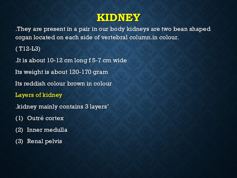
Kidney
The kidneys are bean-shaped organs located on the posterior abdominal wall, one on each side of the vertebral column.
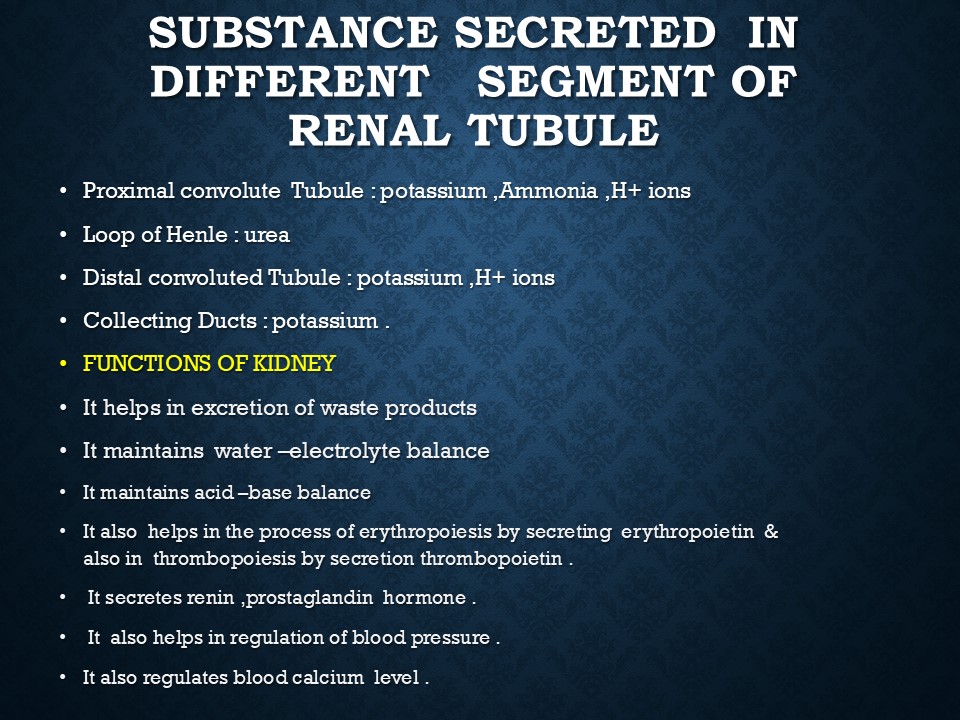
Functions of the Kidneys
1. Excretion of water and waste products of protein metabolism.
2. Excretion of excess salt.
3. Excretion of harmful substances, drugs, and toxins.
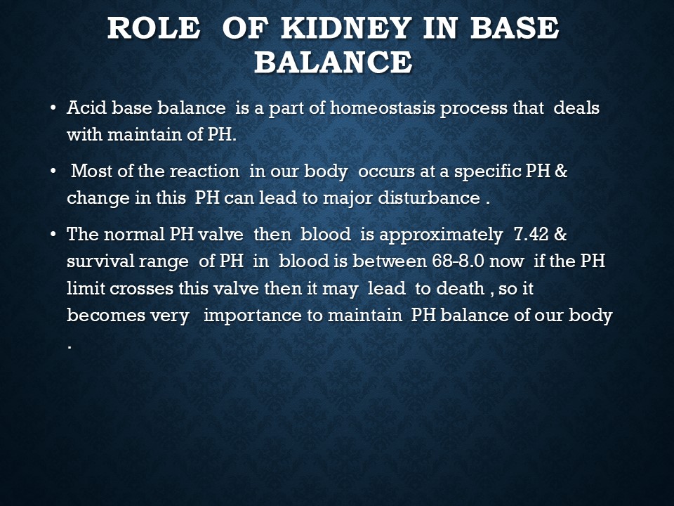
4. Regulation of blood pH.
Position of the Kidneys
The kidneys extend from the last thoracic vertebra (T12) to the third lumbar vertebra (L3). The right kidney is slightly smaller and lower than the left kidney. Each kidney measures 11 cm in length, 5 cm in width, and 3 cm in thickness, with an average weight of 150 grams.
The outer border of the kidney is convex, while the inner border is concave and called the hilum, through which blood vessels, nerves, and the ureter enter and exit. A suprarenal (adrenal) gland is situated on the apex of each kidney.
Structure of the Kidney
Each kidney is covered by a fibrous capsule and consists of:
1. Outer cortex – Reddish-brown in color.
2. Inner medulla – Contains renal pyramids.
3. Renal pelvis – The expanded upper end of the ureter.
Microscopic Structure of the Kidney
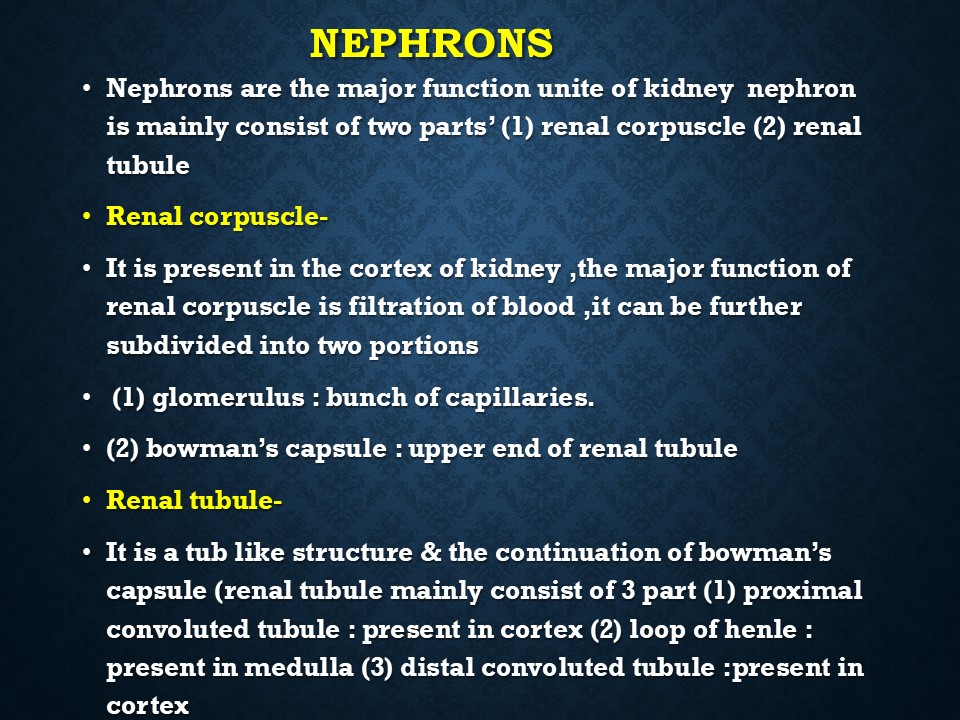
The kidneys contain about one million nephrons, which are the structural and functional units responsible for urine formation.
A nephron consists of:
1. Malpighian bodies, composed of:
Bowman’s capsule – A cup-shaped structure.
Glomerulus – A network of capillaries enclosed in Bowman’s capsule.
2. Renal tubules, divided into:
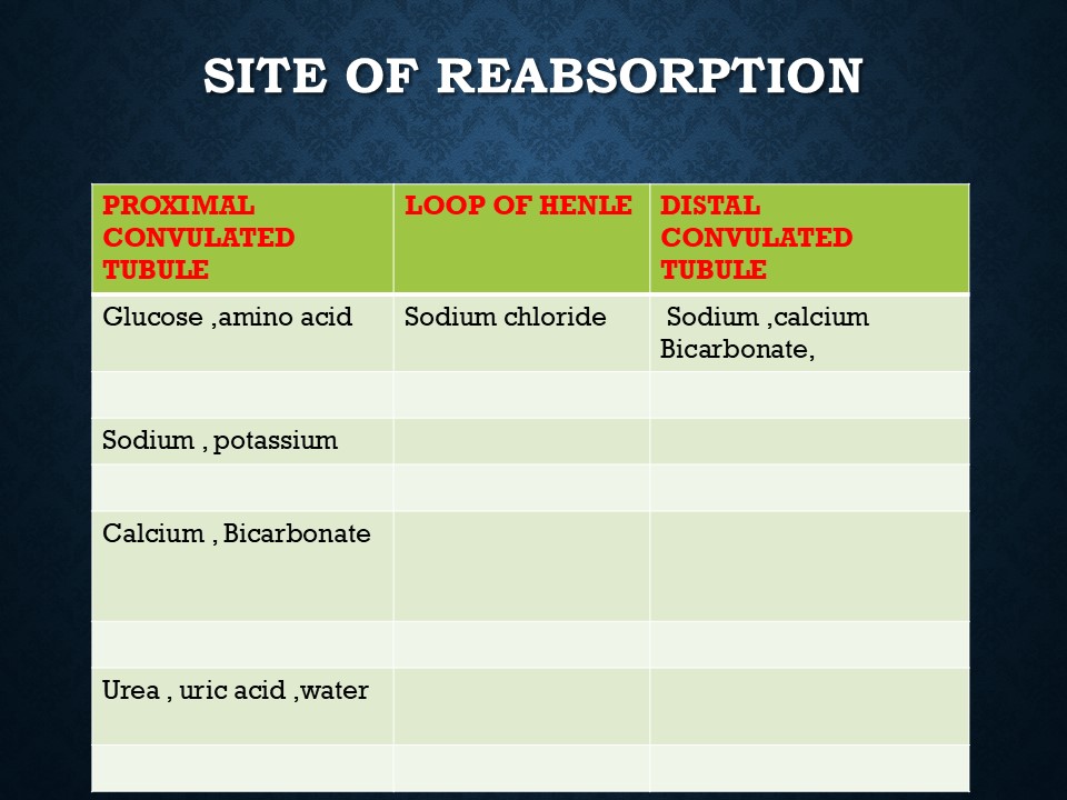
Proximal convoluted tubule – Located in the cortex.
Loop of Henle – Present in the medulla.
Distal convoluted tubule – Present in the cortex.
Collecting ducts – Pass through the medulla and open into the pelvis of the kidney.
Blood Supply to the Kidney
Renal arteries (branches of the abdominal aorta) supply blood to the kidneys.
Renal veins drain deoxygenated blood into the inferior vena cava.
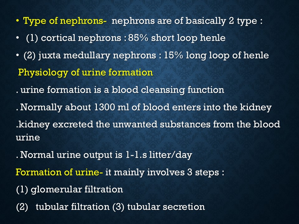
Formation of Urine
Urine formation occurs through three main processes:
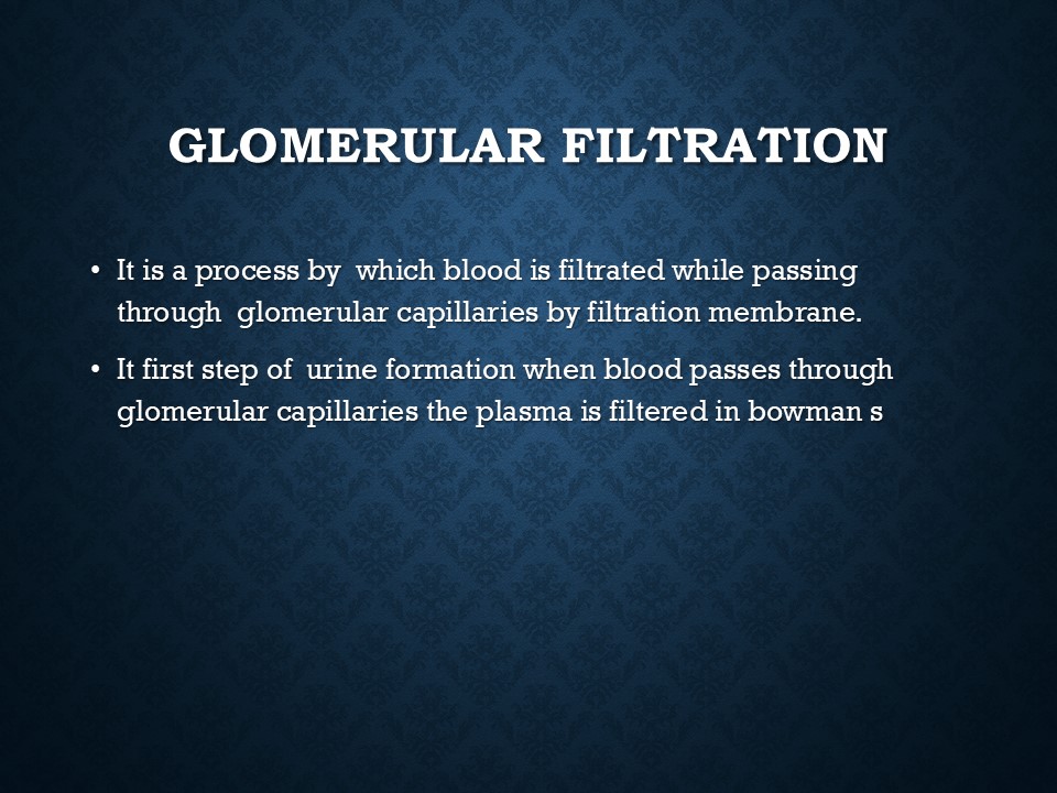
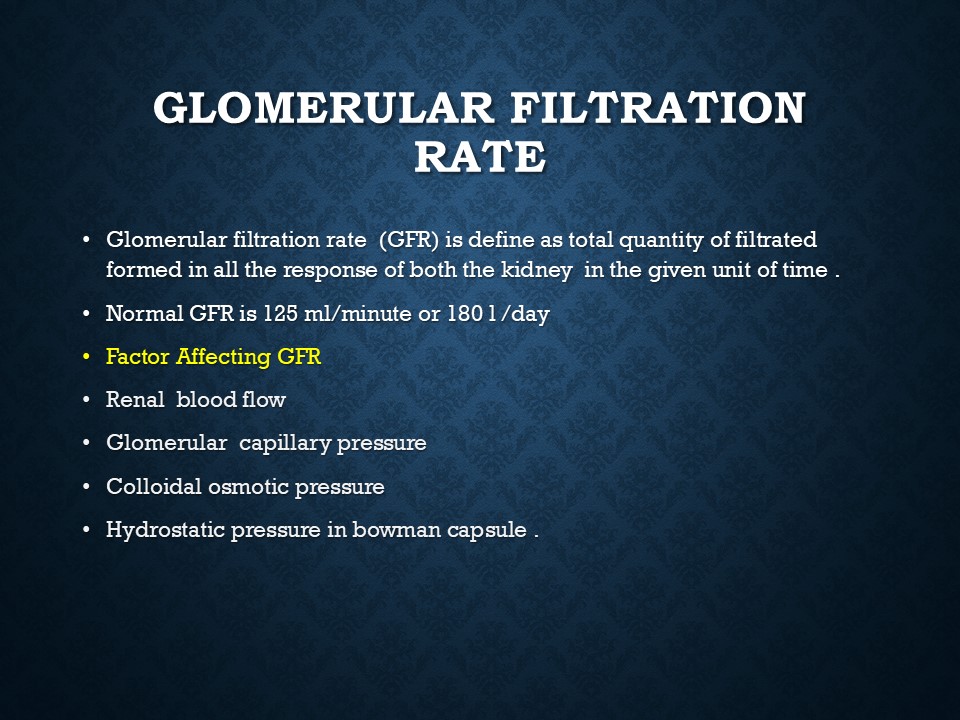
1. Glomerular Filtration
Water, salts, and waste products are filtered in the glomeruli.
The glomerular filtration rate (GFR) is about 100 ml per minute, forming approximately 6 liters of filtrate per hour.
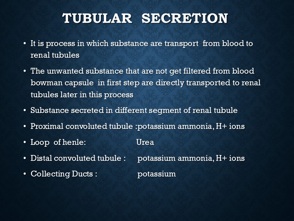
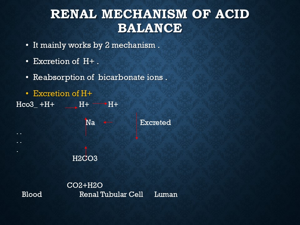
2. Tubular Secretion
Occurs in the renal tubules.
Unwanted substances like potassium, hydrogen ions, and drugs (e.g., penicillin) are actively secreted into the tubule.
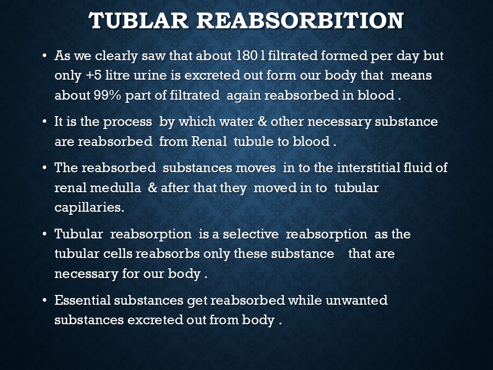
3. Tubular Reabsorption
About 99% of the glomerular filtrate is reabsorbed, reducing urine output to 1.5 liters per day.
Water and essential salts are reabsorbed in the convoluted tubules and collecting duct
The final urine is transported to the renal pelvis, then to the bladder via the ureters.
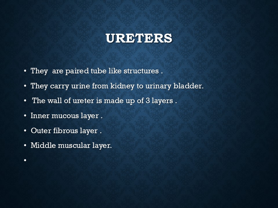
Ureter
A 26 cm long muscular tube that carries urine from the kidney to the bladder.
It has three layers:
1. Outer fibrous layer
2. Middle muscular layer
3. Inner mucosal layer
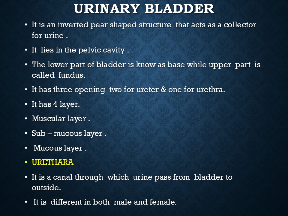
Urinary Bladder
A pear-shaped muscular sac located in the pelvic cavity, behind the pubic symphysis.
It acts as a reservoir for urine.
The trigone of the bladder is a triangular area between the two ureteral openings and the urethral opening.
The bladder has four layers:
1. Serous coat
2. Muscular coat
3. Submucosal coat
4. Mucous membrane (lined by transitional epithelium)
Urethra
The urethra carries urine from the bladder to the outside.
Male Urethra (20 cm long):
1. Pelvic part
2. Perineal part
3. Penile part
Female Urethra (4 cm long):
Begins at the trigone of the bladder and opens anterior to the vaginal opening.
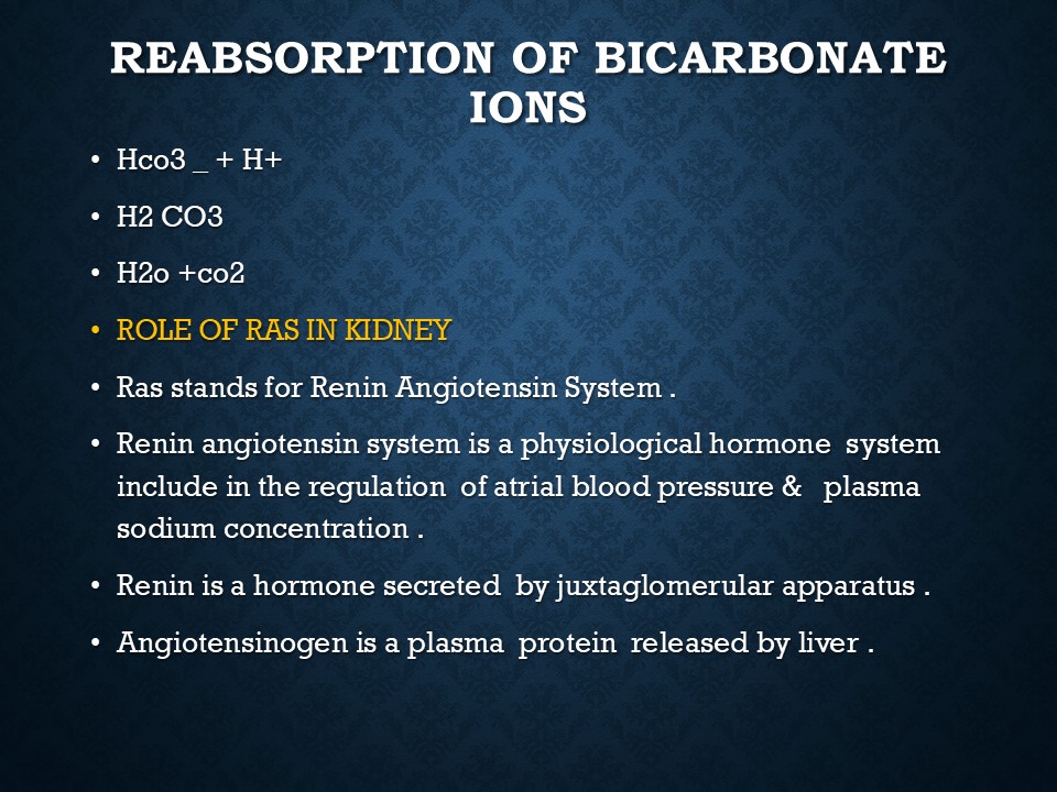
Renin-Angiotensin System
This system regulates sodium and water balance in the body:
1. Renin (from the kidney) is released when renal arterial pressure decreases.
2. Renin converts angiotensinogen to angiotensin I.
3. Angiotensin-converting enzyme (ACE) converts angiotensin I to angiotensin II.
4. Angiotensin II stimulates the adrenal glands to produce aldosterone, which increases salt and water retention.
5. Aldosterone inhibition increases urine output.
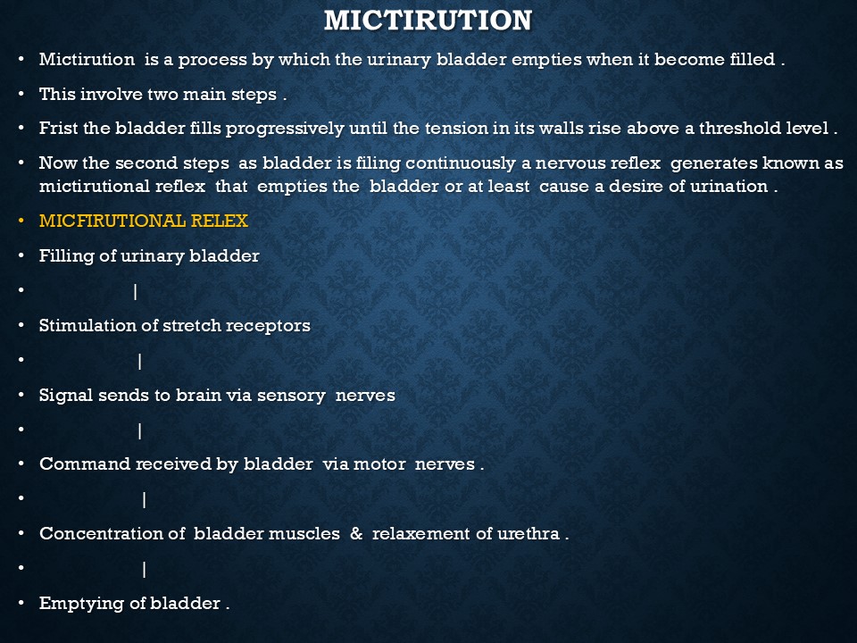
Micturition (Urination Process)
When the bladder fills with 170–230 ml of urine, stretch receptors send signals to the brain.
Bladder contraction and sphincter relaxation result in urination.
Abdominal muscles assist in this process.
Composition of Urine
Volume: 1–2 liters per day
Color: Pale amber
Odor: Aromatic
pH: Slightly acidic (~6)
Specific Gravity: 1.010–1.025
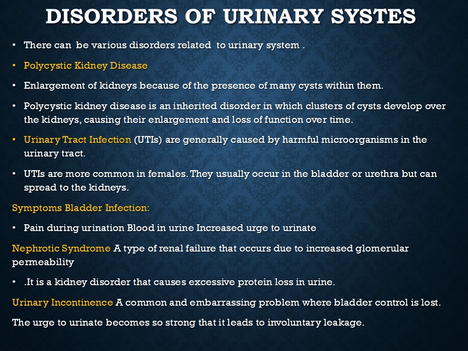
Diseases of the Urinary System
1. Glomerulonephritis – Inflammation of the glomeruli due to infection.
2. Pyelitis – Inflammation of the renal pelvis.
3. Polyuria – Excessive urine production.
4. Anuria – Absence of urine production.
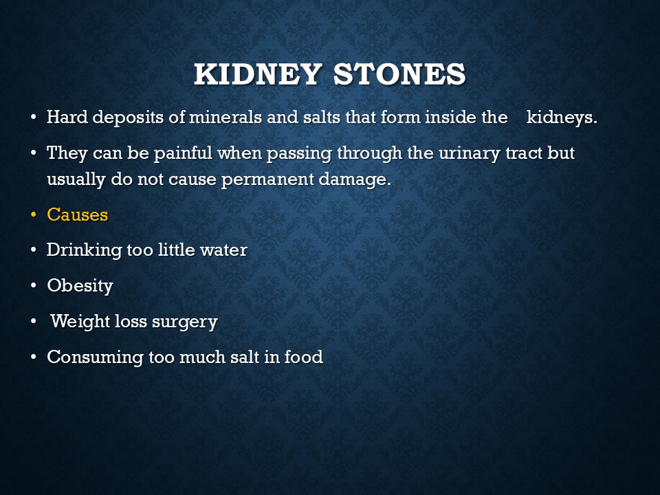
5. Renal calculi (Kidney stones) – Deposits of insoluble substances in the urinary tract.
6. Cystitis – Inflammation of the bladder.
Oedema (Fluid Retention)
Causes of Oedema:
1. Renal failure – Loss of plasma proteins leads to fluid accumulation in tissues.
2. Cardiac oedema – Due to congestive heart failure.
3. Lymphatic obstruction – Can occur after radical mastectomy or elephantiasis (filariasis infection).
4. Thrombosis – Deep vein thrombosis (DVT) in the legs can lead to swelling.
Recommended

Introduction to anatomy & physiology
20 slides • 6 months ago

THE CELL AND CELL THEORY
20 slides • 5 months ago
GENETICS
12 slides • 5 months ago

Digestive system
32 slides • 4 months ago